Friday, April 29, 2016

Brain dissection

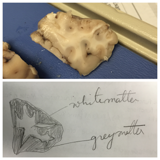
Today in class we dissected a sheep´s brain. We started by observing the outside of the brain and then made our first cut. We made a longitudinal cut first and made a horizontal cut latter just to see the difference between white and grey matter. Here are some pictures/ explanations of this lab.


The Cerebrum's function is to interpret sensory and neural functions relating to voluntary movements.
The cerebellum regulates muscle activity.
The brainstem connects signals from the body and the brain. 
The function of the myelin in a neuron is that it helps the signal flow and it is wrapped around the axon. 
The thalamus helps regulate sleep and sensory input while the hypothalamus maintains homeostasis. 
The optic nerve connects images from the retina to the brain.
The medula oblongata regulates breathing, digestion, sneezing, swallowing, and other vital functions.
Pons is the bridge between the cerebrum and the cerebellum.
The midbrain regulates temperature, vision, hearing, and motor control.
The corpus callousum is the bridge between the right and left hemisphere of the brain. 


Wednesday, April 27, 2016

Eye dissection lab

In our class we dissected a sheep's eye. In the first picture to the left, you can see the external eye. In the middle of the eye is the cornea (which  is a greyish color).  The cornea's function is to protect the eye and it is the first entry point of light. Surrounding the cornea is the sclera which is referred to as "the whites of our eyes". The sclera's function is to protect because it is very tough. On the right of the picture you can see the fatty tissue that we have taken off the eye already.


Here is a picture of the back of the eye. The probe is pointing at the optic nerve. The function of the optic nerve is to transfer signals from the eyes to the brain.


 Next we cut open the eye. In this picture, you can see the lens in the middle of the eye and it is surrounded by aqueous humor. It provides the eye with nutrition and it maintains the internal pressure in the eye.


The lens (pictured below)'s function is to focus on objects to provide the retina with a clear image.


The black/grey ciliary body surrounds the pupil and iris. The function of the ciliary body is to produce aqueous humor fluid. It also keeps the lens in place.

The pupil's function is to let light enter the eye. The iris controls how much light is entering the eye.


This is a picture of the vitreous humor that seeped out of the eye during the dissection. It fills the space behind the lens and it's function is to help the eye maintain its shape.


In this last picture, you can see the tapetum lucid which is a reflective coating that helps the sheep have good night vision. The scalpel in this picture is pointing to the optic disc. The optic disc is where the nerves attach to the optic nerve.


Monday, April 18, 2016

My banana bread recipe

I just got my mom´s banana recipe and I am starting to figure out how much whole wheat flour to substitute in for all purpose flour. According to this website, if you substitute 3/4 cups whole wheat for every cup of all purpose flour it should work out. I might also test out how substituting 1 cup of whole wheat to every cup all purpose flour would affect the recipe. My mom also said in her recipe that you could, optionally, substitute brown sugar for white sugar. I´m debating whether or not to include the effects of that change in my blog post. If I were to mention substituting brown sugar in, I would have to do more research. Researching would take extra time and I would not have enough time to do a second recipe if I wanted to.However, I am leaning towards doing the extra research. 

Sunday, April 17, 2016

Clay brain lab

In class we created models of the brain. We used different colors of clay for different parts of the brain. The main challenge for our group was figuring out how to draw the two hemispheres, but after we figured that out, it was easier to label all the little details.

Wednesday, April 13, 2016

Living without a part of your brain

For homework we read an article called "Woman of 24 found to have no cerebellum in her brain." This woman's case was particularly special because it is extremely rare to live without your cerebellum. In her childhood, it took her years longer to achieve basics skills like walking. The cerebellum controls balance, posture, coordination and speech. The most important take away from this article was how the brain was able to adapt and work even with the missing part.

Would a person survive without their frontal lobe or a damaged frontal lobe? The frontal lobe controls emotions, memory, language, and problem solving. People with damaged frontal lobe suffer from memory loss and they have been associated with risk taking. A person would be able to survive without their frontal lobe, but they would not be able to do much. They would be paralyzed.